

Insulin is the body’s primary anabolic hormone: it unlocks cells so glucose can leave the bloodstream and enter muscle, fat, and liver tissue for storage or energy. According to the Cleveland Clinic, insulin acts like a key that opens the cell door to glucose, and without it, blood sugar climbs to dangerous levels. Beyond glucose, insulin drives fat synthesis, suppresses fat breakdown, and stimulates protein building — making the role of insulin in hormonal regulation far broader than most people realize.
Florvahealth’s educational content on hormonal health draws on this same science, particularly as it applies to women navigating PMS, cycle irregularities, and metabolic symptoms tied to insulin and hormone regulation.
Key Takeaways
Insulin is the body’s central anabolic hormone, regulating glucose, fat, and protein metabolism through a tightly coordinated signaling cascade that, when disrupted, drives conditions from type 2 diabetes to PCOS.
| Point | Details |
|---|---|
| Insulin’s core role | It enables glucose entry into cells and simultaneously drives fat synthesis, protein building, and hepatic glucose suppression. |
| Incretin effect magnitude | GLP-1 and GIP account for roughly 60–70% of postprandial insulin secretion, linking gut health directly to insulin dynamics. |
| Insulin resistance and disease | Insulin resistance is the central lesion in type 2 diabetes and also drives PCOS, dyslipidemia, cardiovascular disease, and fatty liver. |
| Women’s hormonal connection | Estrogen supports insulin sensitivity at physiologic levels; its decline at menopause and dysregulation in PCOS both worsen insulin resistance. |
| Lifestyle leverage | Exercise, sleep quality, meal composition, and gut health all measurably shift insulin sensitivity, often within days. |
Table of Contents
- What is insulin and where does it come from?
- The four primary metabolic functions of insulin
- How does insulin work at the cellular level?
- What raises or lowers insulin secretion?
- How insulin interacts with other hormones
- Insulin in health and disease: resistance, diabetes, and metabolic consequences
- Clinical relevance: therapeutic insulin and daily life implications
- Insulin and women’s hormonal health
- How the body clears and degrades insulin
- How genetic variations affect insulin production and action
- What the evidence on insulin really tells us
- Sources
What is insulin and where does it come from?
Insulin is a peptide hormone built from 51 amino acids. It is produced exclusively by beta (β) cells located in clusters of endocrine tissue called the islets of Langerhans, scattered throughout the pancreas. Those islets are small but densely organized: alpha (α) cells sit alongside β cells and secrete glucagon, while delta (δ) cells produce somatostatin. All three cell types communicate directly with each other through paracrine signaling, which means the hormone output of one cell type immediately influences its neighbors.
After β cells release insulin, it travels first through the portal vein to the liver, where roughly 50% is cleared before it ever reaches general circulation. That hepatic first-pass effect matters clinically: portal insulin concentrations are substantially higher than peripheral concentrations, which is why the liver is the organ most immediately exposed to postprandial insulin surges. Peripheral tissues — muscle and fat — receive a lower but still physiologically meaningful insulin signal.
The four primary metabolic functions of insulin
StatPearls on NCBI Bookshelf summarizes insulin’s metabolic reach across four core functions, each operating in a distinct tissue:
| Function | Primary tissue | Clinical relevance |
|---|---|---|
| Stimulate glucose uptake and glycogen synthesis | Skeletal muscle, adipose | Accounts for ~80% of postprandial glucose disposal |
| Promote lipogenesis and inhibit lipolysis | Adipose tissue, liver | Prevents free fatty acid release; excess drives dyslipidemia |
| Facilitate amino acid uptake and protein synthesis | Skeletal muscle | Drives muscle repair and anabolic growth |
| Suppress hepatic glucose production | Liver | Prevents fasting hyperglycemia between meals |
Glucose uptake and glycogenesis. Skeletal muscle handles the largest share of glucose disposal after a meal. Insulin triggers the translocation of GLUT4 transporters to the muscle cell surface, pulling glucose in. Once inside, glucose is either burned for energy or stored as glycogen. Adipose tissue does the same, though at a smaller scale.

Lipogenesis and lipolysis. In fat cells, insulin simultaneously activates fat synthesis and slams the brakes on fat breakdown. Even a modest rise in insulin is enough to suppress lipolysis, which is why fasting insulin levels matter as much as postprandial peaks. In the liver, insulin promotes de novo lipogenesis — the conversion of excess glucose into fatty acids — which becomes problematic when insulin resistance develops.
Protein synthesis. Insulin increases uptake of amino acids including alanine, arginine, and glutamine into muscle cells, then activates the mTOR pathway to drive protein synthesis. This is why insulin is considered a genuinely anabolic hormone, not just a glucose-lowering one. Muscle maintenance and repair depend on it.
Hepatic glucose suppression. Between meals, the liver continuously releases glucose through glycogenolysis and gluconeogenesis. Insulin suppresses both. When that suppression fails — as it does early in type 2 diabetes — fasting blood glucose rises even before postprandial control collapses.
How does insulin work at the cellular level?
The molecular sequence from insulin binding to glucose entry involves four main steps, and understanding them explains why some drugs work and why resistance develops where it does.
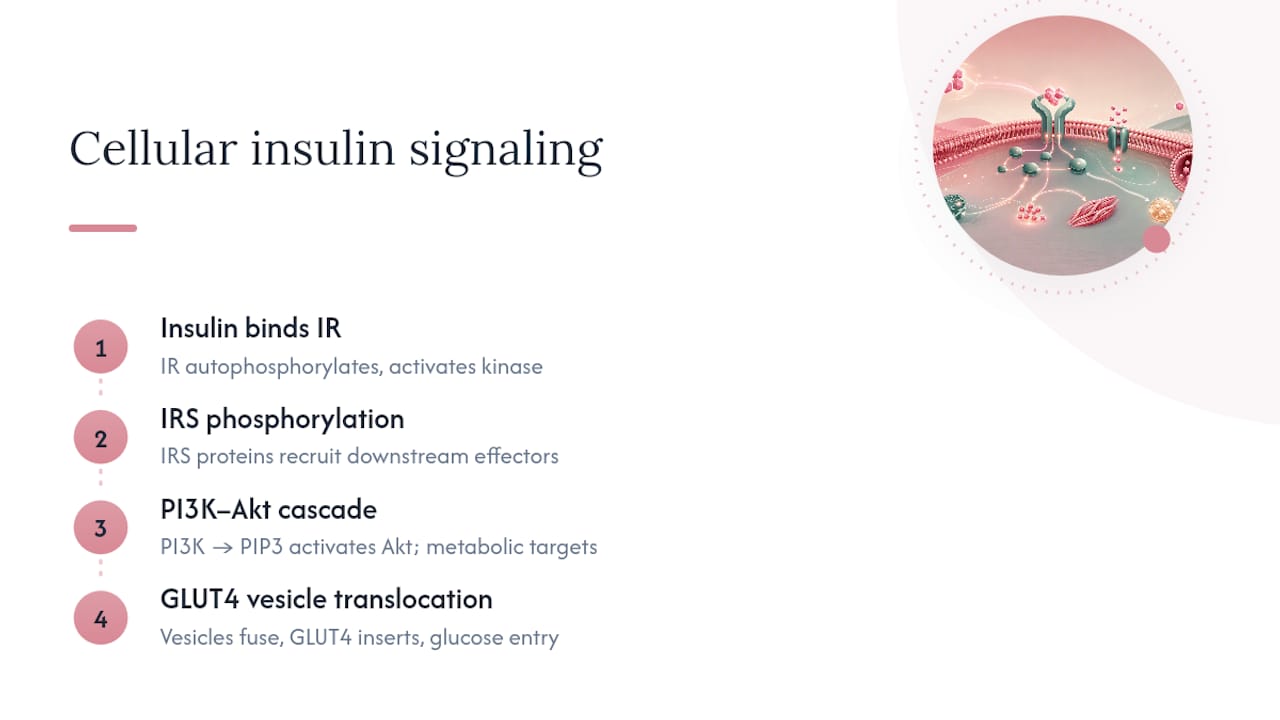
Step 1: Receptor binding. Insulin binds to the insulin receptor (IR), a transmembrane tyrosine kinase receptor present on virtually every cell type. Binding triggers autophosphorylation — the receptor phosphorylates itself on tyrosine residues, activating its kinase function.
Step 2: IRS activation. The activated receptor phosphorylates insulin receptor substrate (IRS) proteins, which serve as docking platforms for downstream signaling molecules.
Step 3: PI3K–Akt cascade. IRS recruits phosphoinositide 3-kinase (PI3K), which generates the lipid messenger PIP3. PIP3 activates Akt (also called PKB, protein kinase B). Akt is the central node: it phosphorylates dozens of downstream targets that collectively drive glucose uptake, glycogen synthesis, lipogenesis, and protein synthesis. A comprehensive PMC review confirms this PI3K–Akt axis as the dominant metabolic arm of insulin signaling.
Step 4: GLUT4 translocation. Akt phosphorylates AS160 (TBC1D4), releasing GLUT4-containing vesicles from intracellular storage. Those vesicles fuse with the plasma membrane, inserting GLUT4 transporters that pull glucose into the cell. In insulin-resistant states, this translocation step is impaired — the receptor may still bind insulin, but the signal never fully reaches the vesicle pool.
A parallel arm, the MAPK pathway, branches off IRS and drives cell growth, proliferation, and gene expression. This arm is less affected in typical insulin resistance, which is why hyperinsulinemia in resistant individuals can still drive abnormal cell proliferation — a factor in conditions like PCOS and certain cancers.
Pathway summary: IR → IRS → PI3K → PIP3 → Akt → AS160 → GLUT4 at membrane → glucose entry.
What raises or lowers insulin secretion?
Glucose is the primary trigger, but insulin secretion is modulated by a surprisingly wide network of signals.
Glucose-stimulated insulin secretion (GSIS). When blood glucose rises, β cells take up glucose via GLUT2 transporters and metabolize it. The resulting rise in the ATP-to-ADP ratio closes ATP-sensitive potassium channels, depolarizing the cell membrane. Voltage-gated calcium channels open, calcium floods in, and that calcium surge triggers exocytosis of insulin-containing granules. This is the core mechanism that sulfonylurea drugs exploit.
The incretin effect. Oral glucose produces a much larger insulin response than the same amount of glucose given intravenously. The difference is the incretin effect: gut hormones GLP-1 (glucagon-like peptide-1) and GIP (glucose-dependent insulinotropic polypeptide) are released from intestinal cells within minutes of eating and amplify β-cell insulin output. According to a PMC review on incretin physiology, the incretin effect accounts for roughly 60–70% of postprandial insulin secretion in healthy individuals. GLP-1 receptor agonists (semaglutide, liraglutide) work by mimicking this gut-islet axis.
Other stimulators and inhibitors:
- Amino acids (especially leucine and arginine) directly stimulate insulin release independent of glucose, which is why a high-protein meal raises insulin even without carbohydrates.
- Fatty acids at physiologic levels potentiate GSIS; chronically elevated free fatty acids impair it.
- Parasympathetic nervous system (vagus nerve) stimulates insulin release in anticipation of meals — the cephalic phase response.
- Sympathetic nervous system and catecholamines (epinephrine, norepinephrine) suppress insulin secretion during stress or exercise, prioritizing glucose availability.
- Somatostatin from δ cells inhibits both insulin and glucagon release, acting as a local brake on islet output.
- Circadian rhythm shapes insulin sensitivity across the day: cells are most insulin-sensitive in the morning and less so in the evening, which is why late-night eating produces a larger glucose excursion for the same meal.
Pro Tip: Eating a small amount of protein and fiber before your carbohydrate-heavy portion of a meal can blunt the postprandial glucose spike and reduce the magnitude of the insulin surge. This is worth discussing with your clinician, especially if you track fasting glucose or have a family history of metabolic disease.
How insulin interacts with other hormones
Insulin does not operate in isolation. A Nature Reviews Molecular Cell Biology paper describes the hormonal system as deliberately asymmetric: insulin is the sole hormone that prevents hyperglycemia, while multiple counter-regulatory hormones prevent hypoglycemia. That asymmetry reflects how dangerous low blood sugar is to the brain.
Counter-regulatory hormones:
- Glucagon (from α cells) is the most immediate counter-regulator. When blood glucose falls, glucagon stimulates hepatic glycogenolysis and gluconeogenesis. Insulin suppresses glucagon release through direct paracrine signaling within the islet — a tight local feedback loop.
- Cortisol raises blood glucose by promoting gluconeogenesis and reducing peripheral glucose uptake. Chronic stress means chronically elevated cortisol, which chronically opposes insulin — a direct pathway from psychological stress to metabolic dysfunction.
- Catecholamines (epinephrine, norepinephrine) mobilize glucose rapidly during acute stress or exercise by stimulating glycogenolysis and suppressing insulin secretion simultaneously.
- Growth hormone promotes lipolysis and reduces glucose uptake in peripheral tissues, opposing insulin’s anabolic effects. This is why growth hormone excess (acromegaly) frequently causes secondary diabetes.
Metabolic hormone cross-talk. Leptin, secreted by fat cells, signals satiety to the hypothalamus and also modulates insulin sensitivity. Adiponectin, another adipokine, enhances insulin signaling in muscle and liver — and adiponectin levels fall as visceral fat accumulates, contributing to insulin resistance. The hypothalamus itself responds to insulin: central insulin signaling suppresses appetite and regulates energy expenditure, which is why insulin resistance in the brain can drive overeating independently of peripheral glucose control.
During fasting, exercise, or stress, glucagon and catecholamines dominate, keeping blood glucose available for the brain and working muscles. Insulin drops to its lowest levels. During recovery from exercise, insulin sensitivity in muscle rises sharply — a window where glucose uptake is more efficient and insulin requirements are lower.

Insulin in health and disease: resistance, diabetes, and metabolic consequences
Insulin resistance means cells require more insulin than normal to achieve the same glucose-lowering effect. The CDC frames insulin resistance as the central metabolic lesion driving type 2 diabetes and a major public-health concern. Initially, the pancreas compensates by secreting more insulin. Over time, β cells exhaust their capacity, insulin output falls relative to demand, and blood glucose rises persistently.
Type 1 vs. type 2 diabetes. Type 1 diabetes is an autoimmune destruction of β cells, resulting in near-total loss of insulin production. Without exogenous insulin, glucose cannot enter cells and the body catabolizes fat and muscle for energy, producing ketones. Type 2 diabetes is a combination of insulin resistance and relative insulin deficiency — β cells are still present but functionally impaired. The distinction matters for treatment: type 1 always requires insulin; type 2 often does not, at least initially.
Downstream consequences of chronic insulin dysfunction:
- Hyperglycemia damages blood vessels and nerves through glycation of proteins and oxidative stress.
- Dyslipidemia follows from impaired suppression of lipolysis: elevated free fatty acids drive hepatic triglyceride synthesis and VLDL secretion.
- Cardiovascular disease is the leading cause of death in people with type 2 diabetes, driven by dyslipidemia, hypertension, and endothelial dysfunction.
- Peripheral neuropathy results from microvascular damage to the nerves supplying the extremities.
- Non-alcoholic fatty liver disease (NAFLD) develops when hepatic lipogenesis outpaces fat export, a direct consequence of insulin resistance in the liver.
According to the CDC, more than 1 in 3 American adults has prediabetes, and the majority are unaware of it — making early recognition of insulin resistance one of the most consequential public-health opportunities available.
Clinical relevance: therapeutic insulin and daily life implications
When exogenous insulin is used. Type 1 diabetes requires insulin from diagnosis. In type 2 diabetes, insulin is added when oral agents and lifestyle changes no longer maintain adequate glucose control, during pregnancy complicated by diabetes, or during acute illness with severe hyperglycemia. Insulin formulations range from rapid-acting analogs (lispro, aspart) that cover meals to long-acting basal insulins (glargine, detemir) that suppress overnight hepatic glucose output.
The most important risk: hypoglycemia. The International Diabetes Federation identifies hypoglycemia as the most common and clinically significant side effect of insulin therapy. Symptoms include shakiness, sweating, confusion, and rapid heartbeat; severe episodes can cause loss of consciousness or seizures. Anyone using insulin needs a clear plan for recognizing and treating low blood sugar, including carrying fast-acting glucose.
Other recognized side effects include weight gain (because insulin promotes fat storage) and injection-site lipohypertrophy from repeated injections in the same location.
Lifestyle factors that genuinely move insulin sensitivity:
- Exercise is the most potent non-pharmacologic lever. Aerobic exercise increases GLUT4 expression in muscle; resistance training builds more muscle mass, expanding the body’s glucose storage capacity. Both effects persist for hours after a session.
- Sleep matters more than most people expect. Even one night of poor sleep measurably reduces insulin sensitivity the following day.
- Diet composition affects insulin dynamics through fiber content, glycemic index, and meal timing. Foods that support hormone balance tend to be those that also moderate postprandial glucose excursions — whole grains, legumes, non-starchy vegetables, and adequate protein.
- Gut health connects to the incretin axis: a diverse gut microbiome supports GLP-1 production, which amplifies insulin secretion appropriately after meals.
Thoughtful snack timing and composition — pairing protein or fat with carbohydrates rather than eating carbohydrates alone — consistently reduces postprandial glucose spikes in research settings.
Insulin and women’s hormonal health
The interaction between insulin and female sex hormones is one of the most clinically underappreciated aspects of metabolic health. Estrogen at physiologic levels generally supports insulin sensitivity by upregulating insulin receptor expression and enhancing PI3K–Akt signaling in muscle and fat. When estrogen levels are dysregulated, that protective effect breaks down.
PCOS. Polycystic ovary syndrome is the most common endocrine disorder in women of reproductive age. Insulin resistance is present in roughly 65–70% of women with PCOS, according to estimates cited in endocrinology literature, and it is mechanistically central: hyperinsulinemia stimulates ovarian androgen production, suppresses sex hormone-binding globulin (SHBG), and raises free testosterone. The result is a self-reinforcing cycle where metabolic dysfunction worsens reproductive symptoms and vice versa. Skin consequences — including hormonal acne — are a direct downstream effect of that androgen excess, a connection explored in Florvahealth’s content on hormonal eczema and skin outcomes.
Women with PCOS who address insulin resistance through diet, exercise, and targeted support often see improvements in cycle regularity and androgen-related symptoms alongside metabolic markers.
Pregnancy. Insulin resistance increases progressively across the second and third trimesters, driven by placental hormones including human placental lactogen and progesterone. This is physiologically normal — it redirects glucose toward the fetus — but in women with limited β-cell reserve, it tips into gestational diabetes. After delivery, insulin sensitivity typically normalizes, though gestational diabetes significantly raises lifetime risk of type 2 diabetes.
Menopause. The decline in estrogen at menopause removes its insulin-sensitizing effect. Postmenopausal women show measurably higher fasting insulin and greater visceral fat accumulation compared to premenopausal women with similar body weight, which partly explains the rise in cardiovascular risk after menopause. Cycle tracking, dietary attention to glycemic load, and targeted wellness support are practical starting points — alongside clinical evaluation for anyone experiencing significant metabolic symptoms.
How the body clears and degrades insulin
Insulin is not a permanent signal. After secretion, it has a half-life of roughly 5–6 minutes in circulation, and the body clears it through two main mechanisms.
Hepatic clearance is the dominant route. The liver extracts approximately 50% of portal insulin on the first pass, and additional clearance occurs with each subsequent hepatic circulation. The primary enzyme responsible is insulin-degrading enzyme (IDE), a zinc metalloprotease that cleaves insulin into inactive fragments. IDE is also expressed in muscle and brain tissue, where it contributes to peripheral insulin clearance and has attracted research interest for its potential role in Alzheimer’s disease (insulin resistance in the brain is increasingly studied as a contributing factor).
Receptor-mediated internalization is the second route. When insulin binds its receptor, the insulin-receptor complex is internalized by endocytosis. Inside the cell, insulin is degraded by IDE and other proteases, while the receptor is either recycled to the membrane or degraded. Chronic hyperinsulinemia accelerates receptor internalization and reduces surface receptor density — one mechanism by which persistently high insulin levels paradoxically reduce insulin sensitivity over time.
Renal clearance accounts for a smaller but clinically relevant fraction, which is why insulin requirements often fall in people with advancing chronic kidney disease as renal degradation capacity declines.
How genetic variations affect insulin production and action
Genetics shapes insulin biology at multiple levels, from production to receptor function to downstream signaling.
Monogenic diabetes. Mutations in single genes can cause diabetes independently of lifestyle. MODY (Maturity-Onset Diabetes of the Young) encompasses at least 14 subtypes caused by mutations in genes including GCK (glucokinase, which sets the β-cell glucose threshold), HNF1A, and HNF4A. MODY is frequently misdiagnosed as type 1 or type 2 diabetes because it presents in young people, but its treatment and prognosis differ substantially. Neonatal diabetes, caused by mutations in KCNJ11 or ABCC8 (the ATP-sensitive potassium channel subunits central to GSIS), can sometimes be treated with sulfonylureas rather than insulin once the genetic cause is identified.
Polygenic risk in type 2 diabetes. Common variants in dozens of genes collectively raise type 2 diabetes risk. Many of the highest-impact variants affect β-cell function rather than insulin action — variants near TCF7L2, for example, reduce incretin-stimulated insulin secretion. Others affect insulin signaling directly: variants in the insulin receptor gene (INSR) cause rare but severe insulin resistance syndromes, while common variants in IRS1 and PPARG modestly reduce insulin sensitivity at the population level.
Insulin gene mutations. Mutations in the INS gene itself can cause misfolded proinsulin that triggers β-cell endoplasmic reticulum stress and progressive β-cell loss — a mechanism distinct from autoimmune type 1 diabetes but with a similar clinical outcome. Genetic testing is increasingly accessible and can clarify diagnosis in atypical presentations, guiding more precise treatment.
What the evidence on insulin really tells us
The science of insulin is mature enough that the core mechanisms are settled, but the clinical application is still catching up in important ways. Most people learn about insulin in the context of diabetes, which frames it as a problem hormone rather than what it actually is: the body’s primary anabolic coordinator, running glucose, fat, and protein metabolism simultaneously.
What gets underestimated is how much insulin biology is modifiable. The incretin effect alone — accounting for 60–70% of postprandial insulin secretion — is directly influenced by what you eat, when you eat it, and the state of your gut microbiome. That is not a small lever. Sleep, exercise timing, and meal composition all shift insulin sensitivity in measurable ways within days, not months.
For women specifically, the estrogen-insulin connection deserves more attention than it typically receives in general health content. Cycle phase, reproductive status, and hormonal transitions all change insulin dynamics in ways that affect energy, skin, appetite, and mood. Treating those symptoms without understanding the metabolic substrate underneath is treating the surface.
If you recognize symptoms that might reflect insulin dysregulation — persistent fatigue after meals, difficulty losing weight despite reasonable effort, irregular cycles, or skin changes tied to your cycle — that is worth a direct conversation with your clinician, not just a lifestyle adjustment. This article is educational context, not medical advice, and clinical evaluation is the appropriate next step for anyone with active symptoms.
Sources
The following sources were used to build this article and are worth consulting directly for deeper reading:
- Insulin — Cleveland Clinic
- Biochemistry, Insulin metabolic effects - StatPearls - NCBI Bookshelf
- About insulin resistance and type 2 diabetes — CDC

Florvahealth’s approach to women’s hormonal health starts with the science, including how insulin, estrogen and gut health interact to shape your cycle, skin and energy. Its probiotics and digestive enzymes support the gut side of that picture. Explore the full Florvahealth range to see how inside-out wellness applies to you.
This article is general information, not a substitute for advice from a qualified doctor. Consult a qualified healthcare professional about your own circumstances before acting on anything here.


